COVID-19 Detection on Chest X-Ray and CT Scan Images Using Multi-image Augmented Deep Learning Model
Abstract
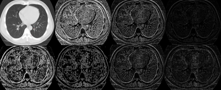
COVID-19 is a deadly and highly infectious pneumonia type disease. RT-PCR is a proven testing methodology for the detection of coronavirus infection in spite of having a lengthy testing time. Sometimes, it gives false-positive results more than the desired rates. To support the conventional RT-PCR methodology or testing independently without RC-PCR methodology for correct clinical diagnosis, COVID-19 testing can be acquired with images of X-Ray and CT Scan of a person. This image-based analysis will make a radical change in the detection of coronavirus in the human body with negligible false-negative and false-positive results. For the detection of COVID-19 in CT Scan and X-Ray images of coronavirus suspected individuals, this paper uses a multi-image augmented Convolutional Neural Network (CNN). For training the CNN model, multi-image augmentation utilizes discontinuity information acquired from the edged images to increase the meaningful examples. With this method, the proposed model exhibits a higher classification accuracy of around 98.97% for X-Ray and 95.38% for CT Scan images. Using multi-image augmentation, X-Ray images achieve a specificity of 98.88% and a sensitivity of 99.07% whereas a specificity of 95.98% and sensitivity of 94.78% are achieved in CT Scan images. The experimental results are also compared with VGG-16 and ResNet-50 models. The evaluation has been performed on publicly available databases comprising chest images of both X-Ray and CT Scan.